RFP Lab
Purpose:
- Make RFP from jelly fish in bacteria
- Learn about steps of genetic engineering.
Materials:
-Materials can be found in the Amgen lab manual
Procedure:
Lab 2A:
Lab 4A:
Lab 5A:
Lab 6A:
Lab 6B:
Data:
Before the 2A Lab:
1. There are two fragments are produced. They are RFP with pBAD and Ara-C with ori with Amp-R. The RFP plus pBAD is 807 BP, and Ara-C, ori, and Amp-R is 4495 BP.
2. We need the RFP gene and Ara-C.
3. The select-able marker allows only the desired bacteria to grow. It separates the bacteria from the desired gene.
2A Questions:
1. Ori = origin of replication; RFP = red fluorescent protein (gene of interest); Amp-R = selectable marker; Ara-C = binds to promoter so we get transcription of gene of interest.
2. Restriction enzymes are a defense mechanism. They cut up foreign bodies.
3. Bacteria retain genes that give them resistance to antibiotics to protect themselves from disease.
4. The central dogma is the same in all organisms.
5. Create a petry dish that grows both Kan and Amp bacteria. Put half of the mixed bacteria into each petry dish and wait a day. The bacteria should have died and then you should be able to separate them apart.
4A Questions:
1. The purpose of growing bacteria that resists ampicillin is to make sure that the desired cell lives.
2. If the bacterial cells are not given arabinose, then the pARA-R plasmid will not turn on the promoter. Then the protein will not turn red.
3. In the LB plate, both the P- and P+ will grow. In the LB/Amp plate, no P- will grow, but P+ will. In the LB/ Amp/Ara plate, there will barely any bacteria growth.
5A Questions:
1. Our predictions sort of matched our results. The LB plate had limited growth. The LB/Amp and LB/Amp/Ara plate matched our prediction.
2. There were no red colonies visible either due to temperature or not enough time in the incubator.
3. The LB-amp-ara plate prevents transcription to occur, which allows RFP to be expressed. The LB-amp does not have Ara.
4. It is important to have multiple copies so there is a greater chance of the promoter being turned on.
5. The RFP gene is expressed as a trait through transcription (central dogma).
6. ???
6A Questions:
1. The red fluorescent protein can be seen separated because of its red cells.
2. The supernatant is clear liquid. The pellet is pink.
6B Questions:
1. Binding Buffer (BB): causes amino acid and protein bind to the resin beads.
Wash Buffer (WB): removes loose proteins that are not bound to the resin beads.
Elution Buffer (EB): takes off protein off resin beads.
Column Equilibration Buffer (CEB): stores resin beads.
2. This time, the supernatant was more pink than clear. The pellet was a little darker pink than the supernatant.
Experimental Overview:
Lab 2A - We verified that we had the correct plasma by using a restriction digest. We cut the plasmid with BamHI and Hind III.
Lab 4A - We verified the plasmid digest by electrophoresis.
Lab 5A - Then we transformed the bacteria with a recombinant plasmid.
Lab 6A & B - The we purified the RFP using chromotography.
Conclusion:
In this lab we attempted to make glowing colonies of bacteria on petri dishes. We did this with restriction enzymes to isolate the RFP gene and then used electrophoresis to make sure it worked. We managed to transfer the gene but the red florescence was missing.
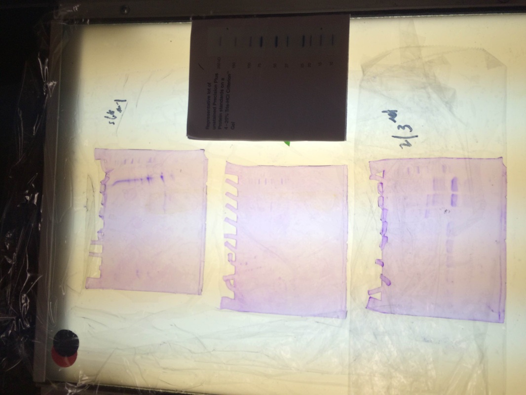
Description: Our particular column was the purple line on the first gel. We had very little protein in the proper places and our gel had some issues as seen in the odd formation. however when looking at 2nd and 3rd period's gels. We can see that some groups had really good RFP extraction. They had a nice thick band at the proper place according to the chart above.
- Make RFP from jelly fish in bacteria
- Learn about steps of genetic engineering.
Materials:
-Materials can be found in the Amgen lab manual
Procedure:
Lab 2A:
- Label two empty microfuge tubes, "R+" and "R-" using a permanent marker
- Add 4.0 ul of 2.5xb, 4.0 pR, and 2.0 ul to the "R+" Tube
- Add 4.0 ul of 2.5xb, 4.0 pR, and 2.0 ul of Distilled Water to the "R-" Tube
- Centrifuge both tubes for four seconds
- Place both tubes into Water Bath and incubate for at least an hour, then remove both tubes and place in a freezer at -20 degrees Celsius
Lab 4A:
- Add 2.0 ul of loading dye to both the "R+" and "R-" tubes
- Microfuge both tubes for several seconds
- Fill electrophoresis box with 1xSB to cover gels
- Dispense 10.0 ul of loading dye, 10.0 ul of "R-", and 10.0 ul of "R+" in individual wells and note their position
- Put lid over box
- Connect the corresponding wires, turn the power on, and set the voltage to 130-135 volts
- After 45-50 minutes, remove gel and examine/photograph
Lab 5A:
- Label 2 empty tubes "P+" and "P-"
- Add 50 ul of competent cells to each tube and keep on ice for remainder of lab
- Add 10 ul of RP to "P+" tube and mix using pipette
- Keep on ice for 15 minutes
- Label each petri dish either LB (Divide in half), LB/Amp (Divide in half), or LB/Amp/Ara (leave whole)
- Carry "P+" and "P-" tubes to 42 degrees Celsius water bath and place them for exactly 45 seconds
- Place back on ice for 1-2 minutes
- Add 150 ul of LB to each tube and gently flick to mix
- Let sit at room temperature for 15 minutes
- Pipet 50 ul of "P-" cells to half of the LB and LB/Amp dishes
- Spread cells using steile spreader
- Repeat for "P+" cells
- Allow dishes to sit right-side up for five minutes and then tape together
- Flip upside-down and let rest in 37 degrees Celsius water bath and incubate for 24-36 hours
Lab 6A:
- Record color of EC tube
- Spin EC tube in microcentrifuge for 5 minutes
- Remove 200 ul of supernatant from EC tube
- Add 1,000 ul of LB/Amp/Ara E. Coli to EC tube
- Repeat steps 2 and 3
- Remove as much supernatant from EC tube as possible
- Add 150 ul of EB to EC tube
- Drag tube across tube rack to resuspend cells
- Add 150 ul of LyB to EC tube and resuspend
- Incubate cells at room temperature overnight
- Dispose of used tips and tubes
Lab 6B:
- Label 2 clean microfuge tubes "SUPER" and "RFP"
- Set liquid waste container underneath stopcock
- Open column and allow liquid to drain into waste container
- Close valve when about 1-2 mm of liquid are left above resin bed
- Microcentrifuge EC tube for about 5 minutes
- Using the P-1000 micropipette, remove 200 ul of supernatant and dispense into "SUPER" tube
- Add 200 ul of BB to the "SUPER" tube and mix gently
- Add 400 ul of "SUPER" tube to the chromatography tube
- Open valve and allow column to drain, but close the valve when 1-2mm of liquid is left above resin bed
- Add 1,000 ul of WB down the side of the column
- Open the valve and allow the column to drain, but close valve when 1-2mm of liquid is left above resin bed.
- Add 2,000 ul of EB down the side of the column
- Set the "RFP" tube under stopcock and allow only the red substance into the tube
- Add 2,000 ul of CEB to column and cap tightly
- Pour waste collection down the drain
- Compare color between other groups
Data:
Before the 2A Lab:
1. There are two fragments are produced. They are RFP with pBAD and Ara-C with ori with Amp-R. The RFP plus pBAD is 807 BP, and Ara-C, ori, and Amp-R is 4495 BP.
2. We need the RFP gene and Ara-C.
3. The select-able marker allows only the desired bacteria to grow. It separates the bacteria from the desired gene.
2A Questions:
1. Ori = origin of replication; RFP = red fluorescent protein (gene of interest); Amp-R = selectable marker; Ara-C = binds to promoter so we get transcription of gene of interest.
2. Restriction enzymes are a defense mechanism. They cut up foreign bodies.
3. Bacteria retain genes that give them resistance to antibiotics to protect themselves from disease.
4. The central dogma is the same in all organisms.
5. Create a petry dish that grows both Kan and Amp bacteria. Put half of the mixed bacteria into each petry dish and wait a day. The bacteria should have died and then you should be able to separate them apart.
4A Questions:
1. The purpose of growing bacteria that resists ampicillin is to make sure that the desired cell lives.
2. If the bacterial cells are not given arabinose, then the pARA-R plasmid will not turn on the promoter. Then the protein will not turn red.
3. In the LB plate, both the P- and P+ will grow. In the LB/Amp plate, no P- will grow, but P+ will. In the LB/ Amp/Ara plate, there will barely any bacteria growth.
5A Questions:
1. Our predictions sort of matched our results. The LB plate had limited growth. The LB/Amp and LB/Amp/Ara plate matched our prediction.
2. There were no red colonies visible either due to temperature or not enough time in the incubator.
3. The LB-amp-ara plate prevents transcription to occur, which allows RFP to be expressed. The LB-amp does not have Ara.
4. It is important to have multiple copies so there is a greater chance of the promoter being turned on.
5. The RFP gene is expressed as a trait through transcription (central dogma).
6. ???
6A Questions:
1. The red fluorescent protein can be seen separated because of its red cells.
2. The supernatant is clear liquid. The pellet is pink.
6B Questions:
1. Binding Buffer (BB): causes amino acid and protein bind to the resin beads.
Wash Buffer (WB): removes loose proteins that are not bound to the resin beads.
Elution Buffer (EB): takes off protein off resin beads.
Column Equilibration Buffer (CEB): stores resin beads.
2. This time, the supernatant was more pink than clear. The pellet was a little darker pink than the supernatant.
Experimental Overview:
Lab 2A - We verified that we had the correct plasma by using a restriction digest. We cut the plasmid with BamHI and Hind III.
Lab 4A - We verified the plasmid digest by electrophoresis.
Lab 5A - Then we transformed the bacteria with a recombinant plasmid.
Lab 6A & B - The we purified the RFP using chromotography.
Conclusion:
In this lab we attempted to make glowing colonies of bacteria on petri dishes. We did this with restriction enzymes to isolate the RFP gene and then used electrophoresis to make sure it worked. We managed to transfer the gene but the red florescence was missing.
Description: Our particular column was the purple line on the first gel. We had very little protein in the proper places and our gel had some issues as seen in the odd formation. however when looking at 2nd and 3rd period's gels. We can see that some groups had really good RFP extraction. They had a nice thick band at the proper place according to the chart above.
Reflection: During this lab our group worked ver
